A new PET tracer that can spot clots anywhere in the body from a single scan, a wearable stocking designed to monitor deep vein thrombosis continuously and a UK trial showing that artificial intelligence still has some way to go before it can be trusted alone. Blood clots remain a strikingly common and often silent killer, and the technology built to catch them earlier is progressing unevenly, but it is progressing.
Venous thromboembolism, the umbrella term for deep vein thrombosis (DVT) and the pulmonary embolisms (PE) that can follow when a clot breaks loose and travels to the lungs, is one of medicine’s more frustrating problems. It is common, it is frequently preventable and it is still missed often enough to kill. Up to two thirds of DVT cases show no symptoms at all before they progress. In the UK, hospital-acquired VTE accounts for thousands of deaths a year, and NHS Digital figures show that 9,030 people died from a VTE-related event within 90 days of hospital discharge in 2019/20, a number that spiked to 11,336 during the first full pandemic year before falling back as care pathways recovered. Globally, pulmonary embolism is the third most common cause of cardiovascular death after stroke and heart attack, and in the United States alone the Centers for Disease Control and Prevention estimates that VTE, DVT and PE combined, affects up to 900,000 people every year.
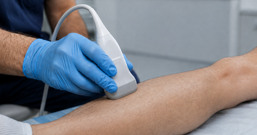
The core diagnostic challenge has not changed much in decades. Compression ultrasound, the standard test for suspected DVT, works well but depends on a trained operator, usually a radiologist or sonographer, and access to one quickly is not always available, particularly out of hours or outside major hospitals. Three separate strands of technology now under development are approaching that bottleneck from different angles: better imaging, artificial intelligence at the point of care and continuous wearable monitoring. Only one of the three has so far been tested rigorously enough to show where its limits lie.
A Single Scan for Clots Anywhere in the Body
At the Society of Nuclear Medicine and Molecular Imaging’s 2026 Annual Meeting in June, researchers from Asan Medical Center in Seoul presented data on a radiotracer called 18F-GP1, which is designed to visualise blood clots directly rather than inferring their presence from indirect structural changes, which is how conventional ultrasound and CT scans work. The tracer targets activated platelets, the cells that cluster to form a clot, allowing PET imaging to pick out a thrombus even in locations that are difficult to assess with standard techniques.
In the study, 46 symptomatic patients underwent 18F-GP1 PET/CT scans that were read independently by three blinded nuclear medicine physicians. The tracer showed high diagnostic accuracy for clots in both the thigh and the calf, as well as a high detection rate for pulmonary embolism occurring alongside DVT, all from a single whole-body scan. No drug-related adverse events were recorded. The work was selected as the SNMMI’s 2026 Henry N. Wagner, Jr. Image of the Year, chosen from nearly 1,500 abstracts submitted to the meeting. Sangwon Han of Asan Medical Center, who led the study, said the approach could allow “a single whole-body PET scan” to evaluate clots in the legs and lungs simultaneously, reducing the need for multiple separate tests. Giuseppe Esposito, chair of the SNMMI’s Scientific Program Committee, suggested the tracer could eventually serve as a platform technology extending beyond DVT into stroke and cardiovascular disease detection more broadly. The tracer has already completed Phase 2 evaluation in DVT, stroke and cardiovascular disease; researchers estimate that with successful Phase 3 trials, it could enter routine clinical practice within five to ten years.
When AI Alone Is Not Yet Good Enough
Not every technology under trial has cleared its bar, and one of the more instructive results of the past year came from a UK study that showed exactly where the limits of artificial intelligence currently sit. AutoDVT, a software tool designed to guide non-radiology staff through a compression ultrasound scan and flag suspected clots, was tested in a multicentre, double-blind study across eleven UK hospitals and published in NEJM AI. Of 294 patients analysed, AutoDVT achieved a sensitivity of 68% and a specificity of 80% when used on its own, well short of the study’s pre-defined targets of 90% sensitivity and 60% specificity. Its negative predictive value was a reassuring 95%, meaning a negative result could largely be trusted, but its positive predictive value was just 28%, meaning a positive flag from the software alone was wrong more often than it was right.
Crucially, when the same scans were reviewed remotely by a clinician alongside the AI output, sensitivity rose to 85%. The researchers also found that 81% of initial scans had to be repeated, a median of three times, because of failed scanning attempts. Their conclusion was blunt: AutoDVT’s accuracy was not yet sufficient for safe standalone use by non-expert operators, and further software optimisation is needed before it can be deployed without a clinician checking its output. It is a useful corrective to the assumption that AI diagnostic tools are ready to be handed to any member of staff. The evidence here suggests they currently work best as an aid to a trained eye rather than a replacement for one.
A Wearable Built to Watch Continuously
The third approach abandons the single-scan model altogether in favour of continuous monitoring. ThrombUS+, a project launched in January 2024 and funded with €9.5 million over three and a half years through the EU’s Horizon Europe programme, is developing a wearable, worn as a stocking or legging, that combines ultrasound sensors, electrical impedance plethysmography and light reflection rheography to track the lower limb continuously and flag early signs of clot formation without requiring an operator to be present.
The project brings together 18 partners across Greece, where the effort is led by the Athena Research Center, alongside teams in Lithuania, France, Germany, Italy, Finland, Spain and the United States. Thorsten Prinz of VDE, the German engineering association involved in the project’s regulatory work, has pointed to the scale of the technical challenge: miniaturising the sensor components without degrading the clinical data they produce, while also satisfying the EU’s Medical Device Regulation and its newer AI Act, which sets minimum standards for trustworthy AI systems in healthcare. The intended users are patients considered at high risk of DVT, including those recovering from surgery, cancer patients, people who are bedridden for extended periods and women during and after pregnancy. The technology is still working through its clinical validation studies, and unlike AutoDVT it has not yet published results against a reference standard, so its real-world performance remains to be seen.
A Field Moving at Different Speeds
Taken together, these three projects show a field advancing unevenly, which is arguably more honest than a single dramatic breakthrough would be. The PET tracer is the most mature of the three, built on established nuclear medicine techniques and already through Phase 2 testing. The wearable is the most ambitious in concept, promising to catch clots before symptoms appear at all, but it remains several years from clinical evidence. AutoDVT sits in between: proven enough to have been tested properly in a real multicentre trial, and honest enough, in the way its results were reported, to show that the technology is not there yet.
That last point matters as much as any individual result. Diagnostic tools for a condition as common and consequential as VTE will only earn a place in routine NHS or hospital practice if their limitations are established as rigorously as their strengths, and the AutoDVT trial is a reminder that AI-assisted diagnosis, for all its promise, still needs a clinician in the loop for now.
Sources include NHS Digital, the Nuffield Trust, NICE, the Society of Nuclear Medicine and Molecular Imaging, News-Medical, NEJM AI, 2 Minute Medicine and VDE (Verband der Elektrotechnik Elektronik Informationstechnik).